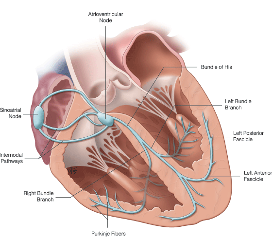
Email : kimberly@avancelearning.com


  Contact : 813-508-1511

All posts by Kimberly

Paced Rhythms

Pacemakers

Pacemakers can be temporary or permanent. Temporary are used in emergencies and usually used only a few days at most.

What does a pacemaker do to an EKG rhythm strip?

it creates pacer spikes.

pacer spike example

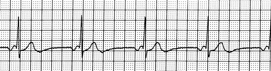
a. Types of Pacing Response- Each pacer has settings programmed into it and is either triggered, inhibited, or both.

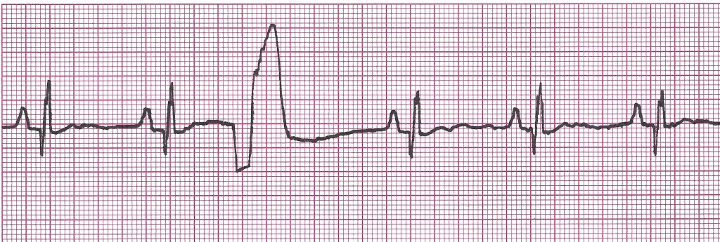
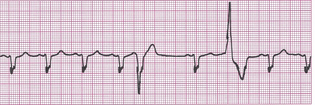
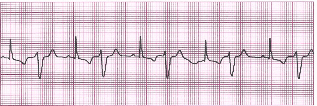
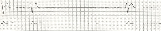
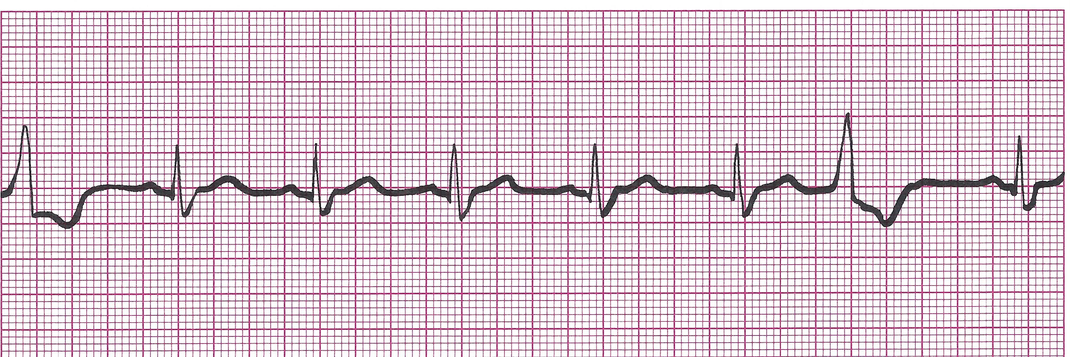
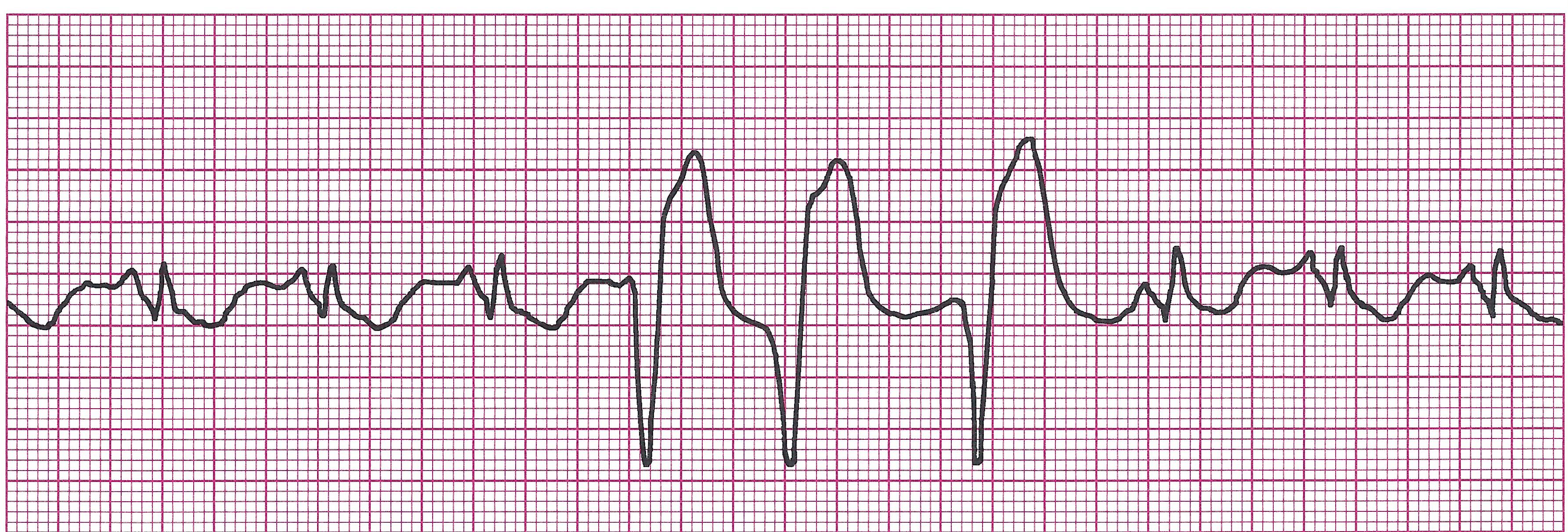
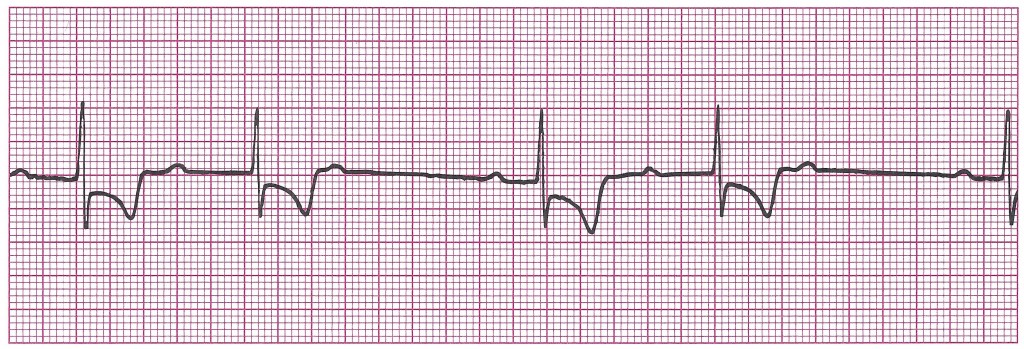
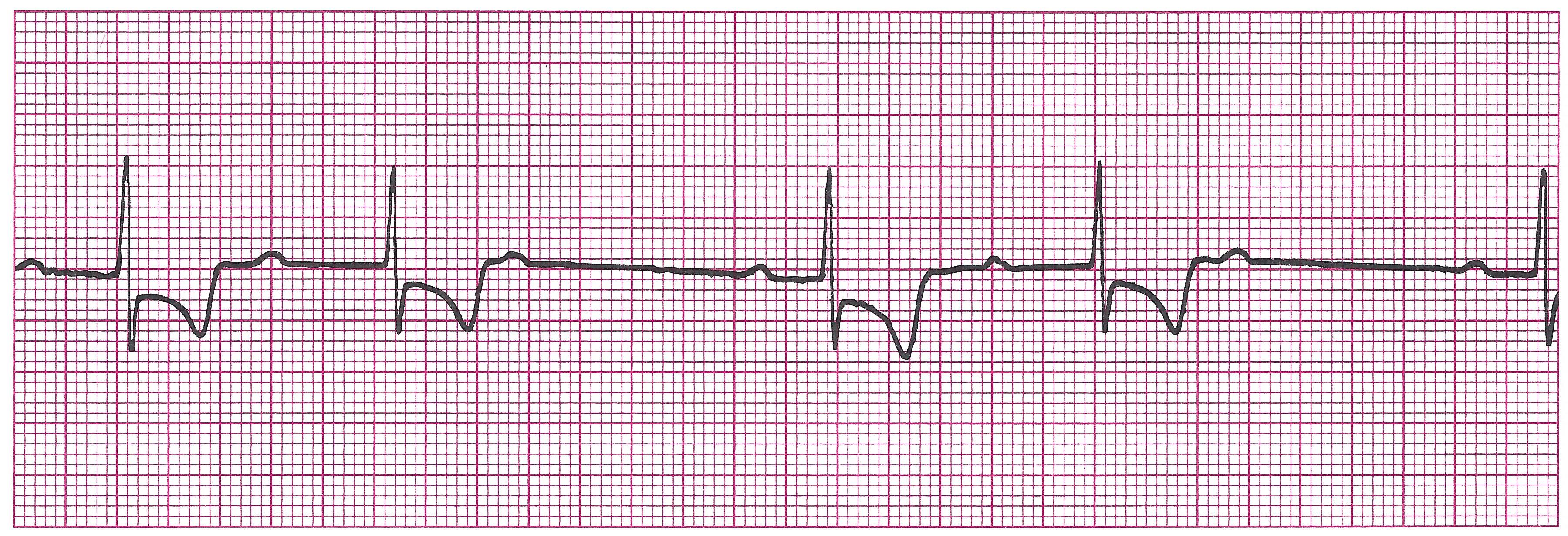
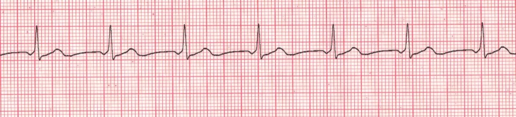
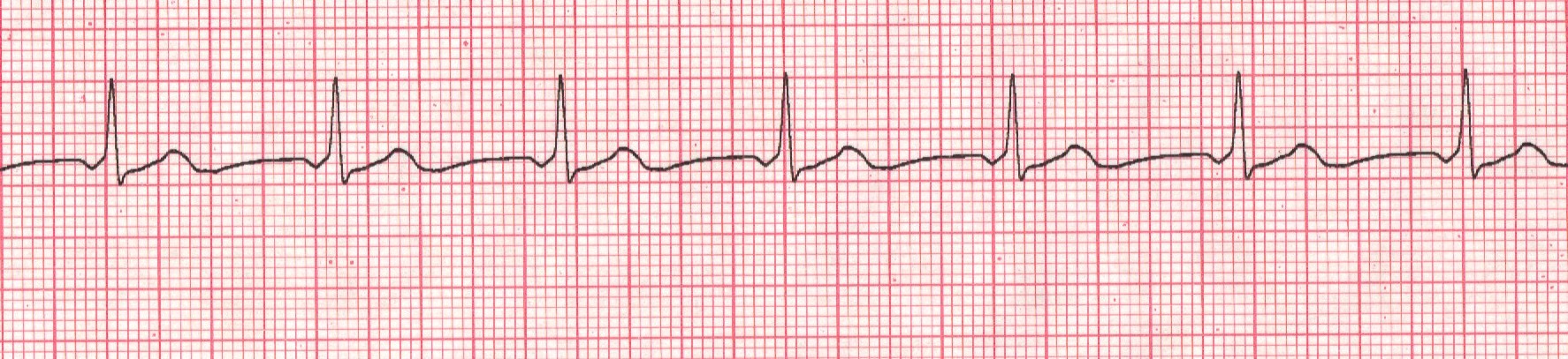
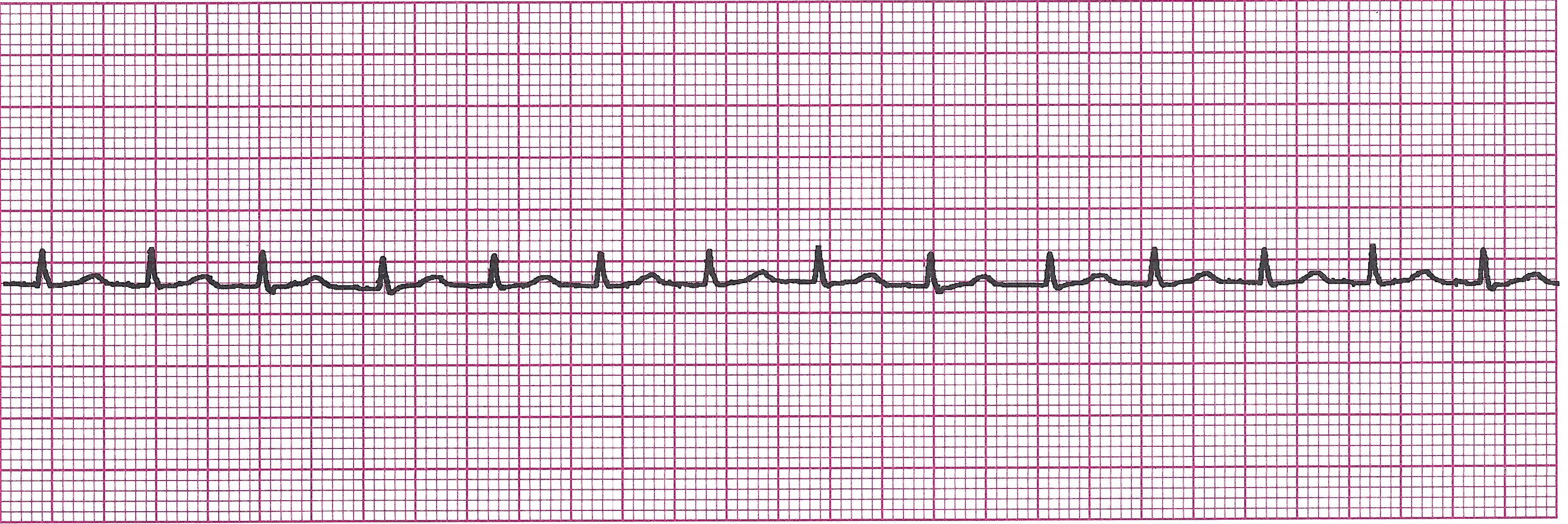
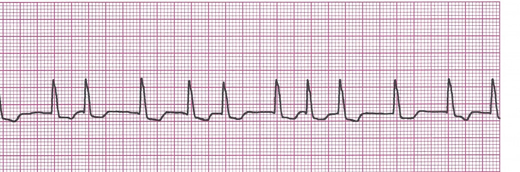
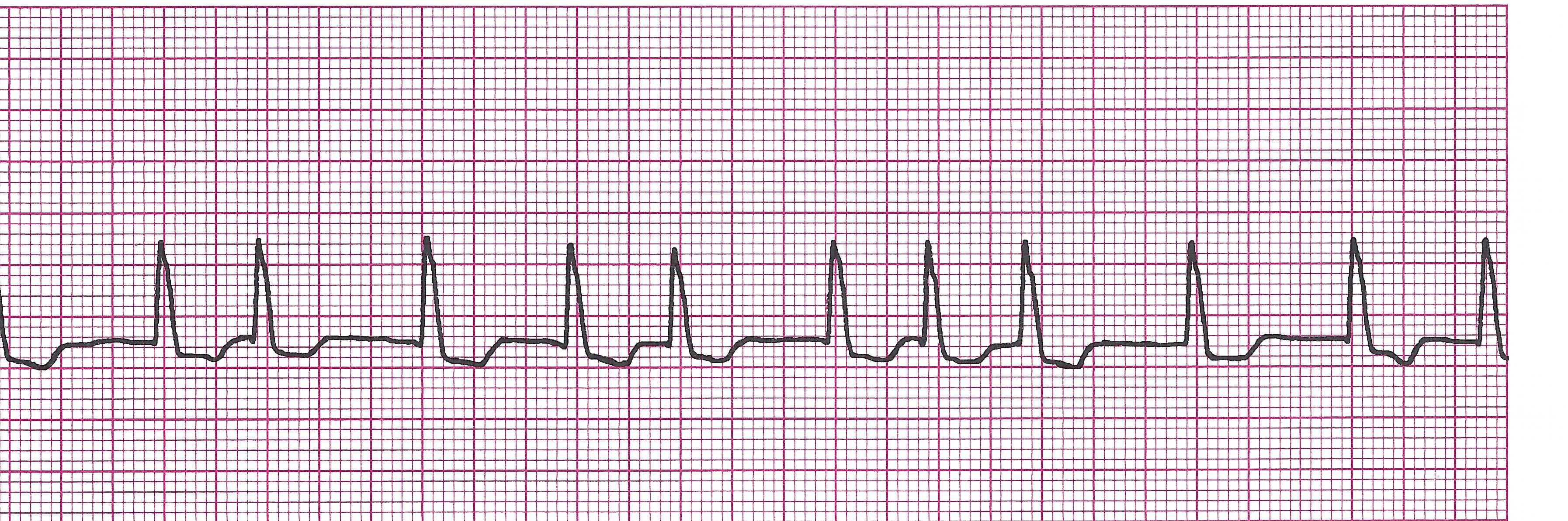
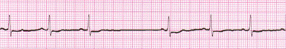
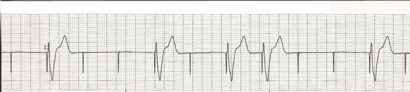
1) Triggered- Fixed rate. Fires according to a predetermined program regardless of patient’s underlying heart rate or rhythm. (i.e. pacer is programmed to send 75 impulses to the ventricles every minute regardless of what the atria are doing as in the strip below).

Example EKG strip with Triggered pacing

(12-27) vent paced 1

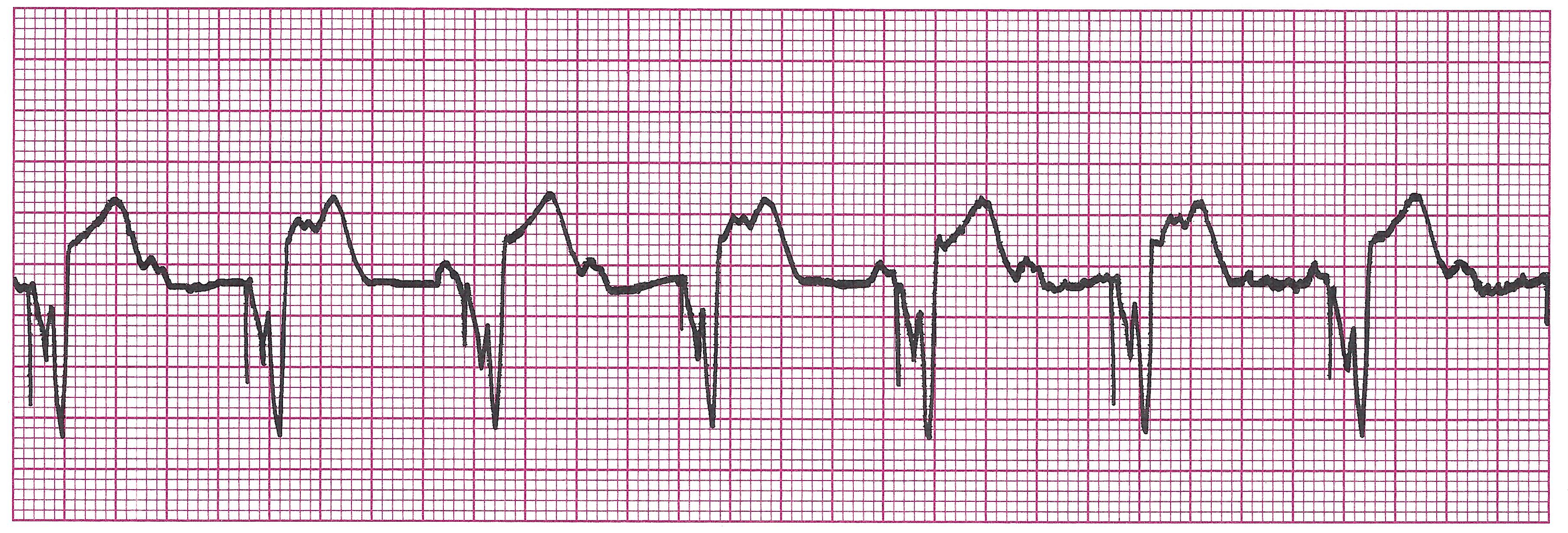
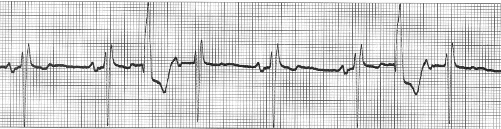
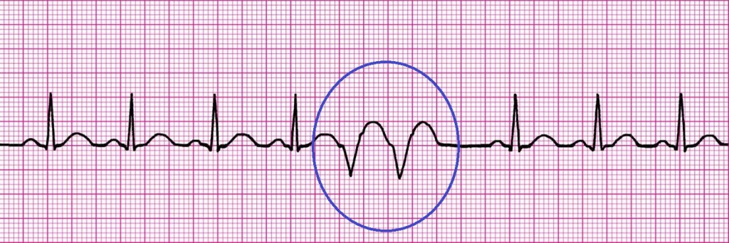
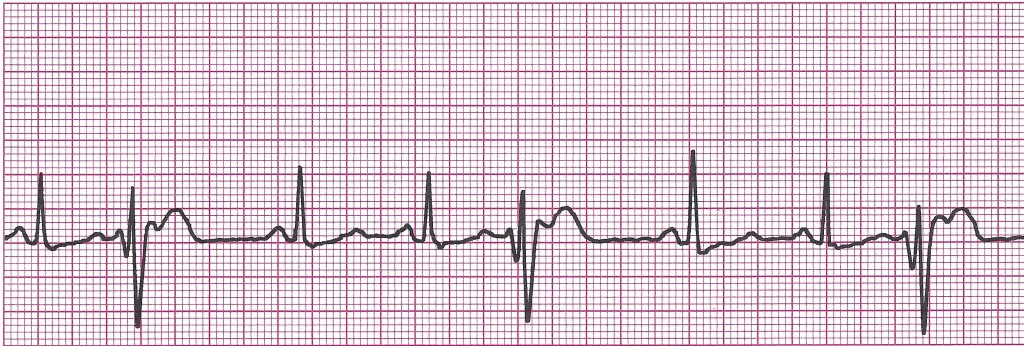
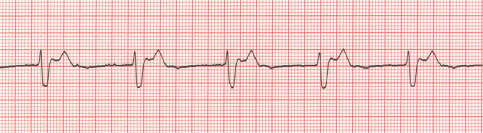
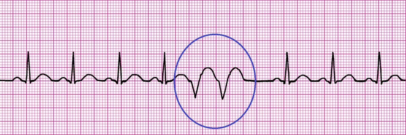
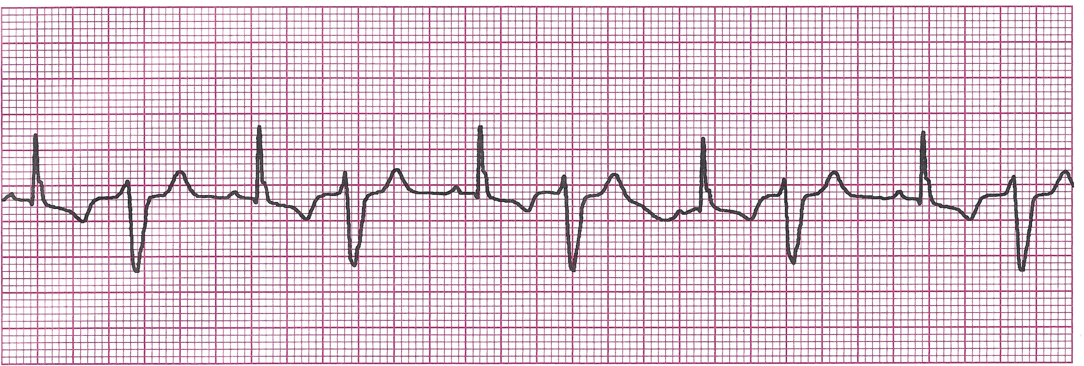
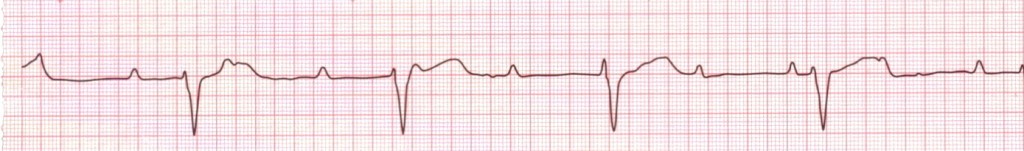
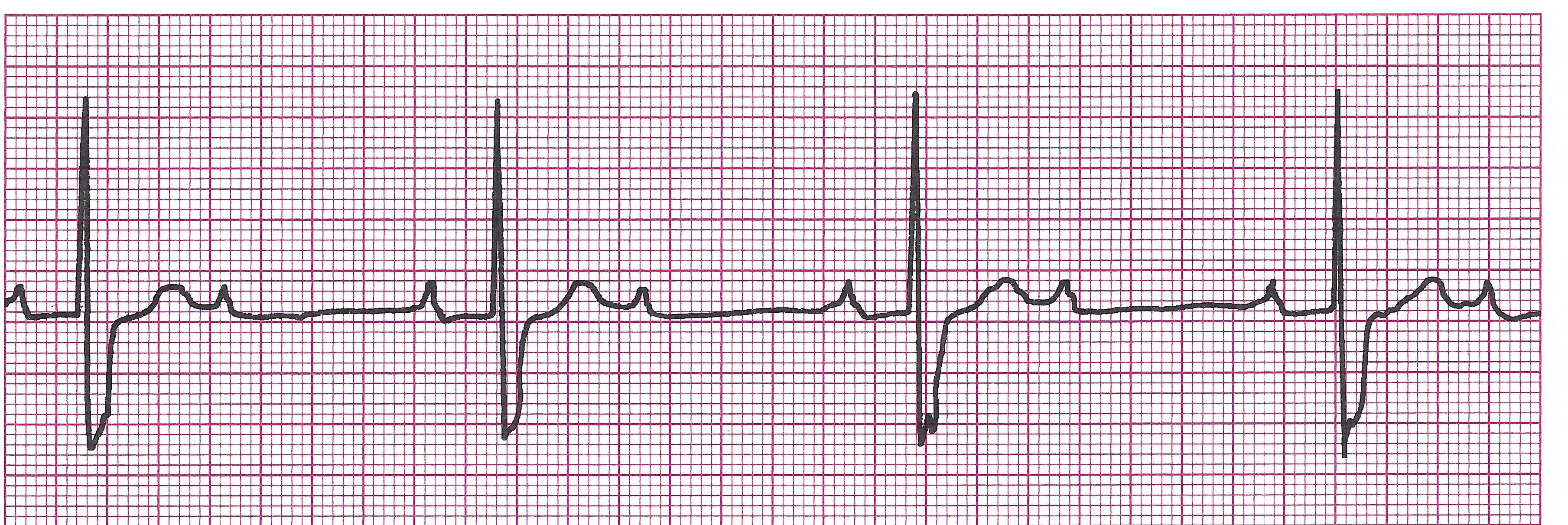
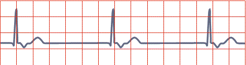
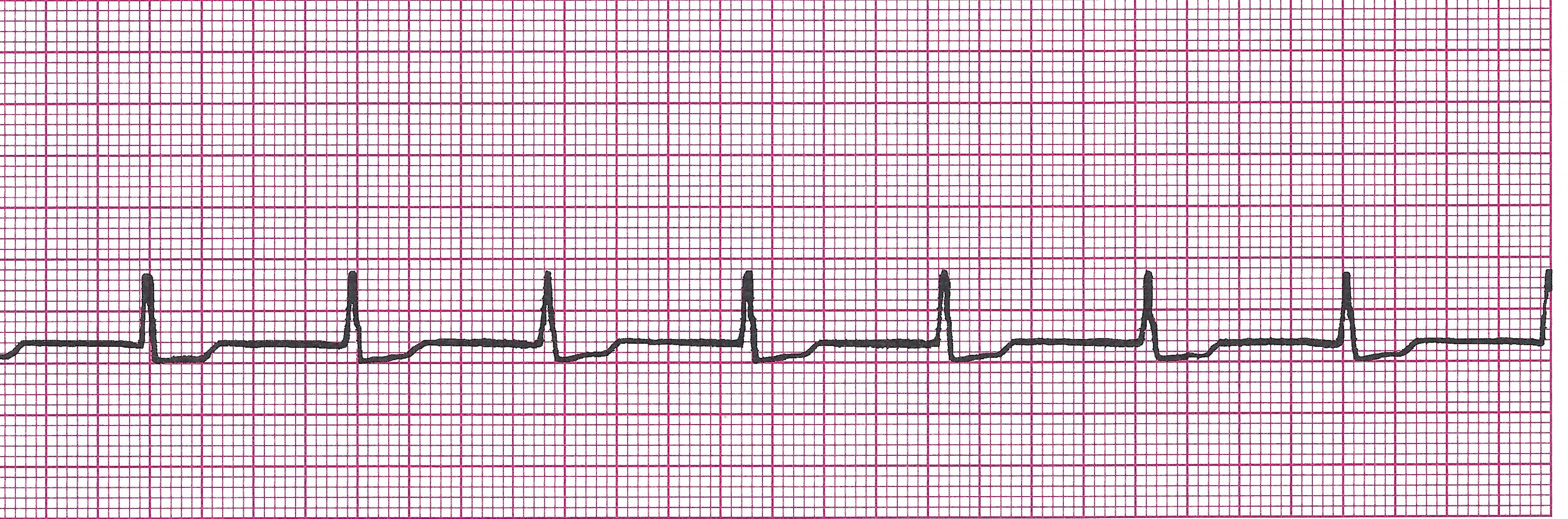
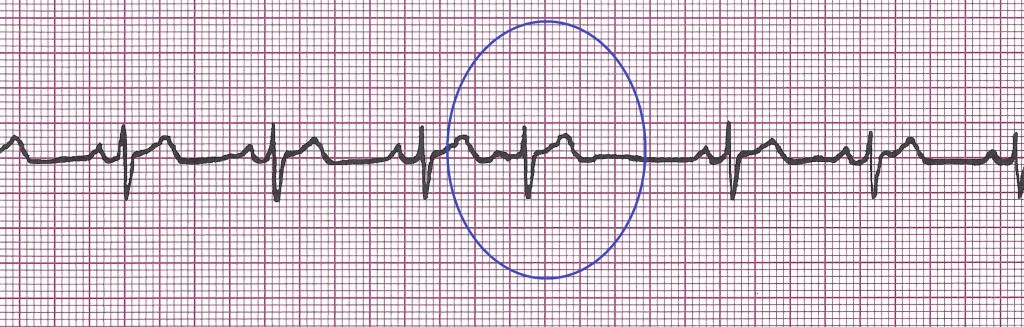
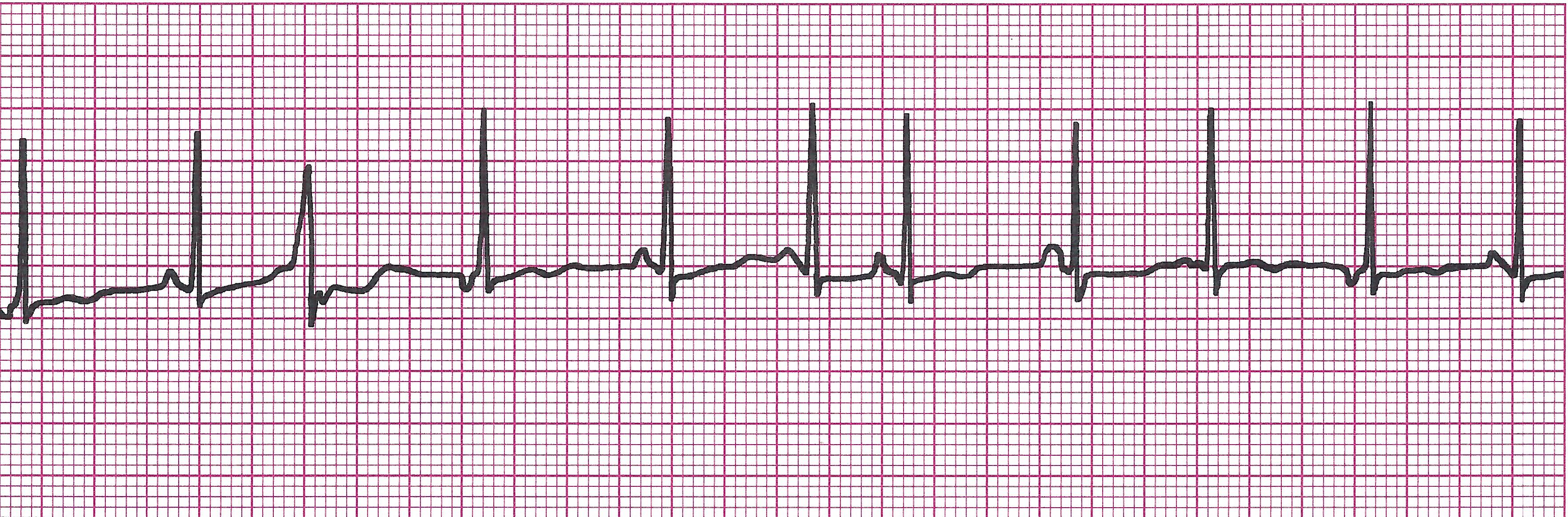
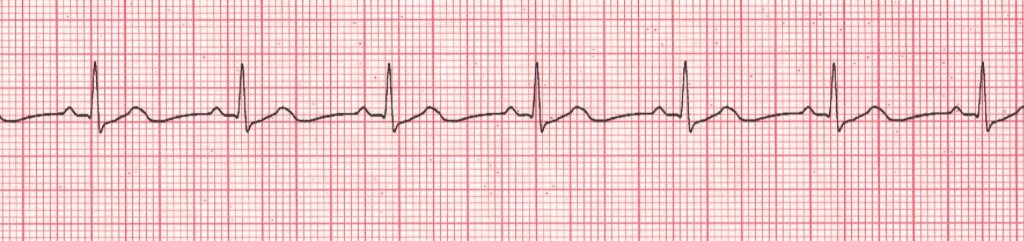
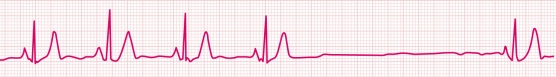
2) Inhibited- Fires only when needed. Senses patient’s own rhythm and inhibits its impulse until needed (i.e. the ventricle or atria stop beating or are beating too slow, etc). In the strip below, you can see the pacer only kicks in after the patient's ventricles fail to beat.

Example EKG strip with Inhibited pacing

demand paced 2

Can be both: Triggered atrial, (this means the pacer sends an impulse to the atria regardless of how the atria are beating) and inhibited ventricular (this means it senses ventricular beats and fires only when ventricles need it to).

b. Common Kinds of Inhibited Pacers

1) Single chamber- Senses and stimulates either atria or ventricles but not both

2) Dual chamber-Works on both chambers simultaneously. Most common is DDD-Dual pacing for both chambers, Dual chamber activity sensing, and Dual response (triggering and inhibition).

c. Assessment

1) Things to assess

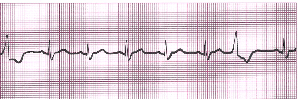
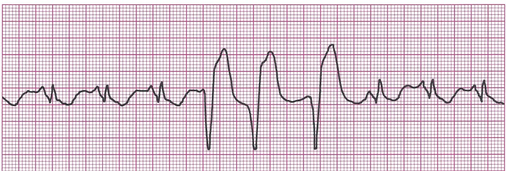
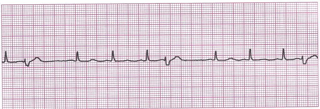
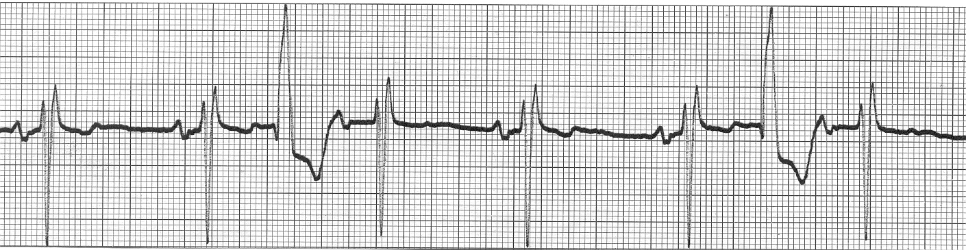
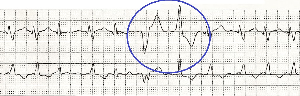
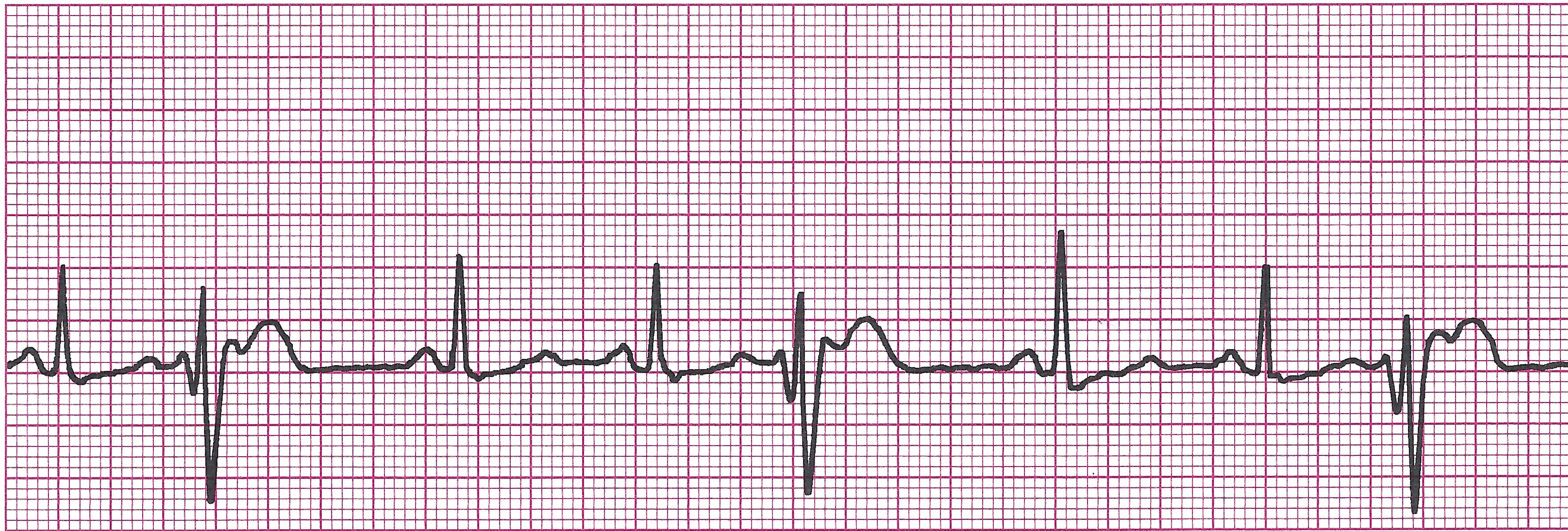
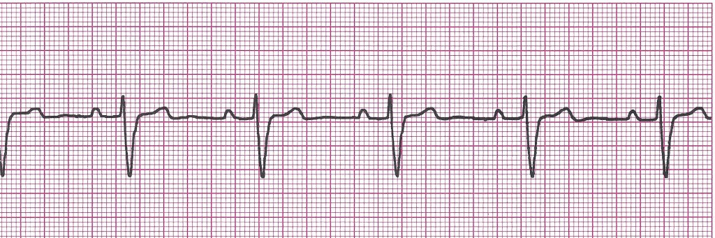
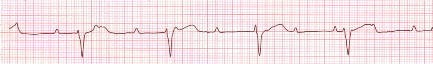
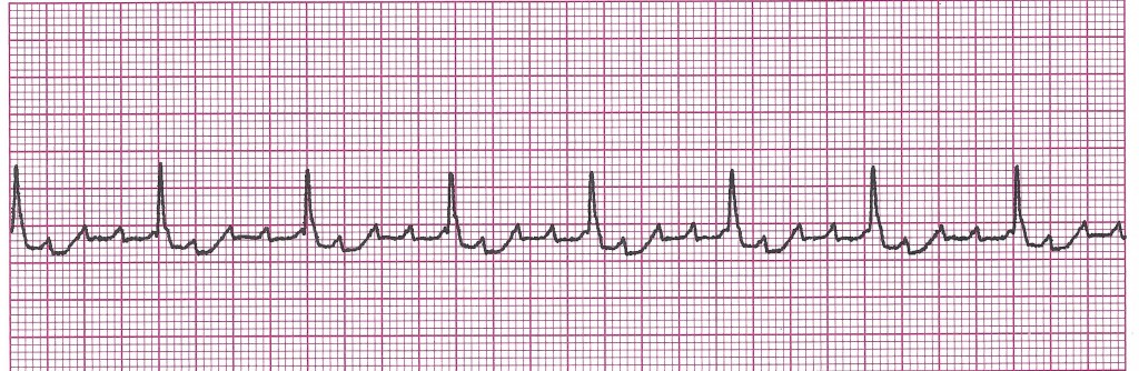
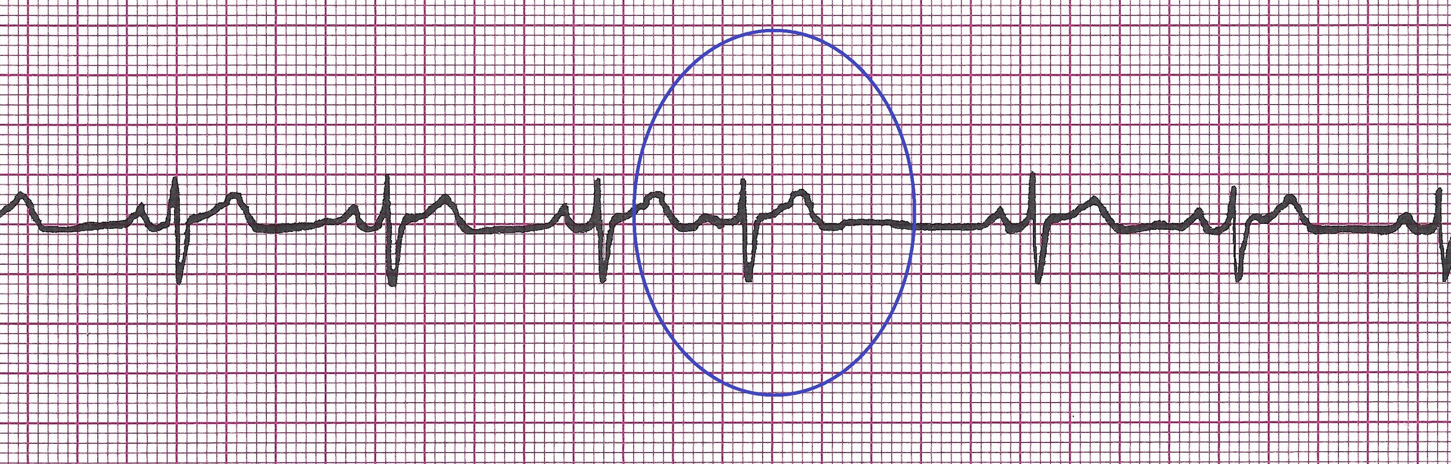
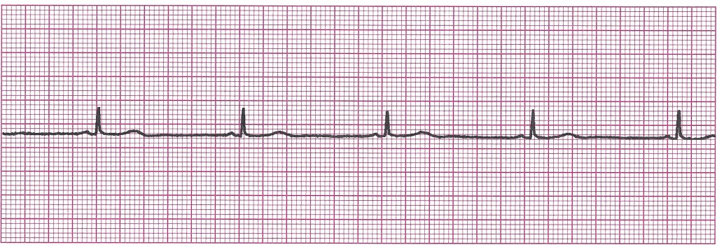
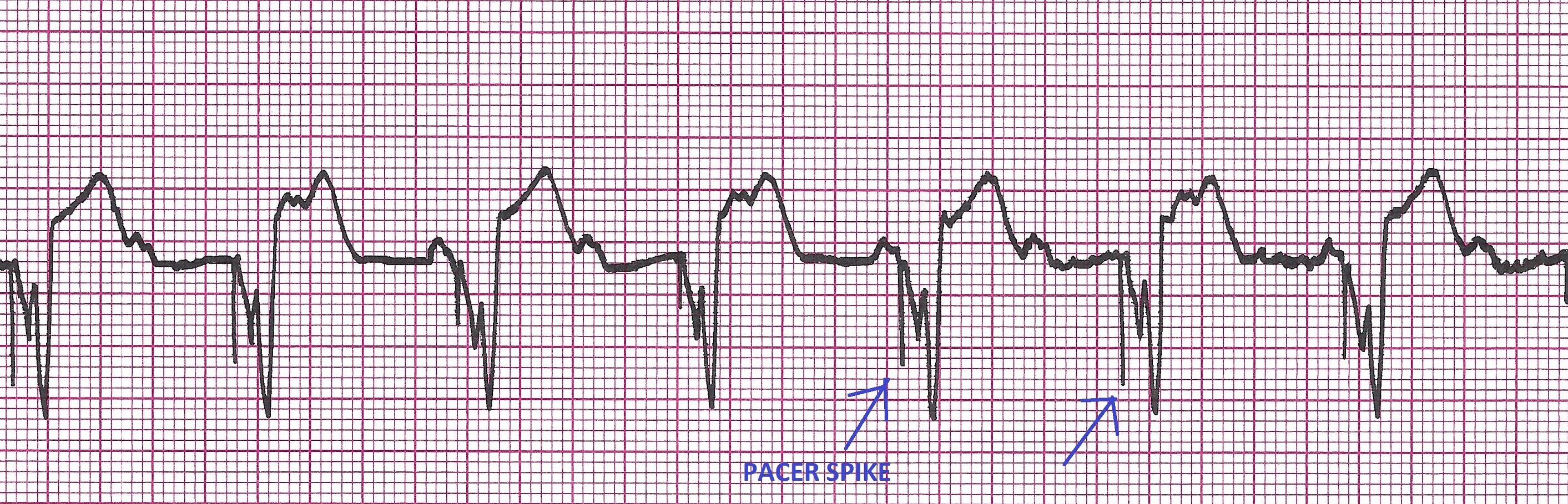
Does each pacer spike capture and get followed by either a P wave or QRS (depending on the type of pacer)?

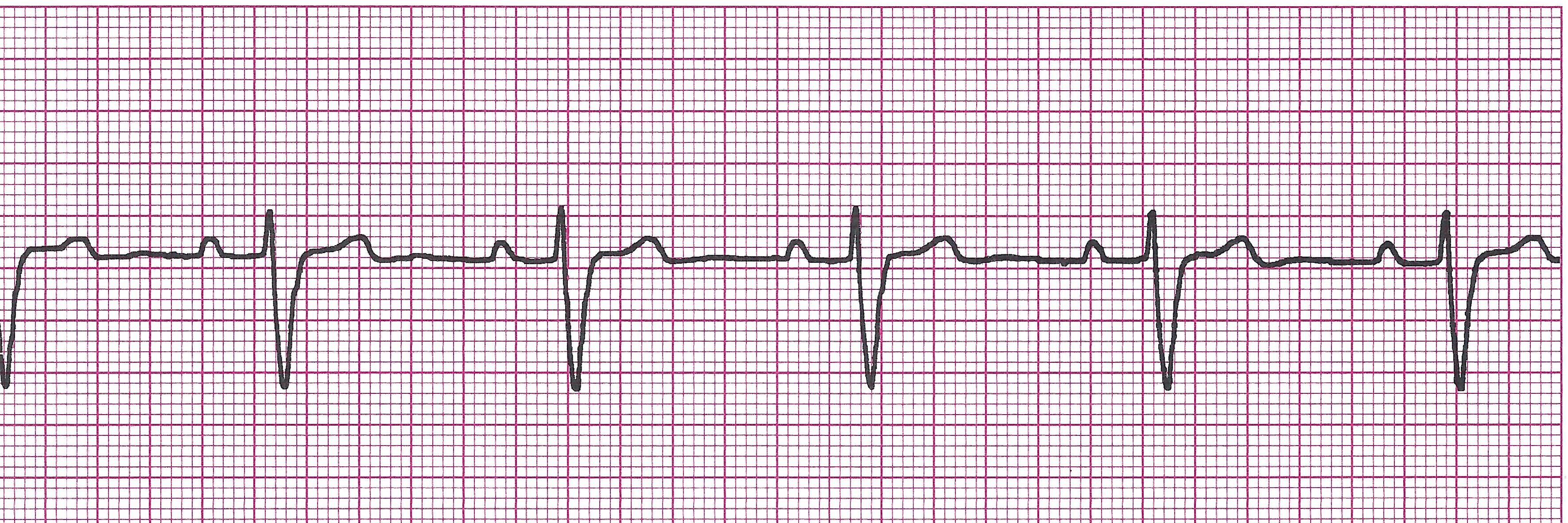
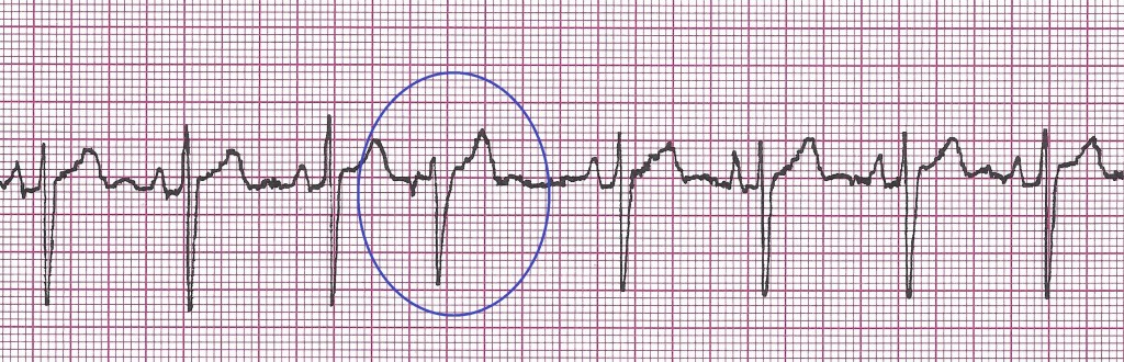
(In the picture below, there are way more pacer spikes than QRS's, not every pacer spike is followed by a QRS meaning that even though the pacer is firing as programmed, the heart is not responding appropriately.)

failure_to_capture

· Is the rate reasonable? (Usually 60-100)

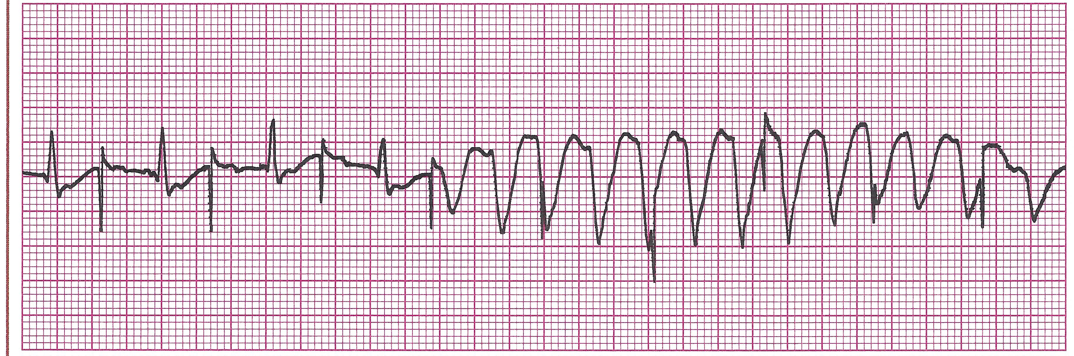
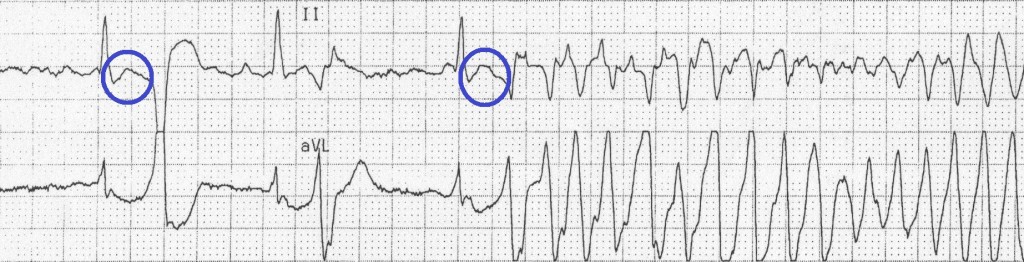
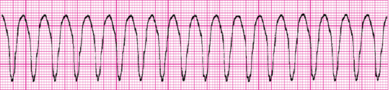
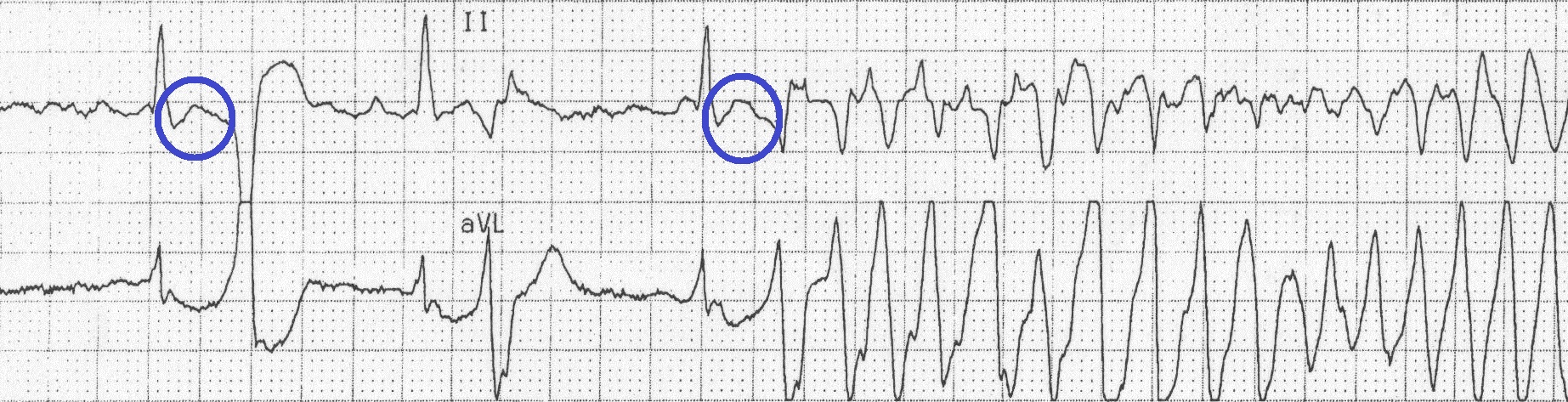
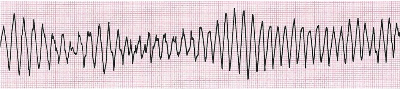
· Are the pacer spikes competing with the patient’s underlying rhythm or falling near T waves? (Spikes falling near T waves can throw a patient into Vtach or Torsades)

· Is pacer firing consistently and reliably?

d. Common Malfunctions

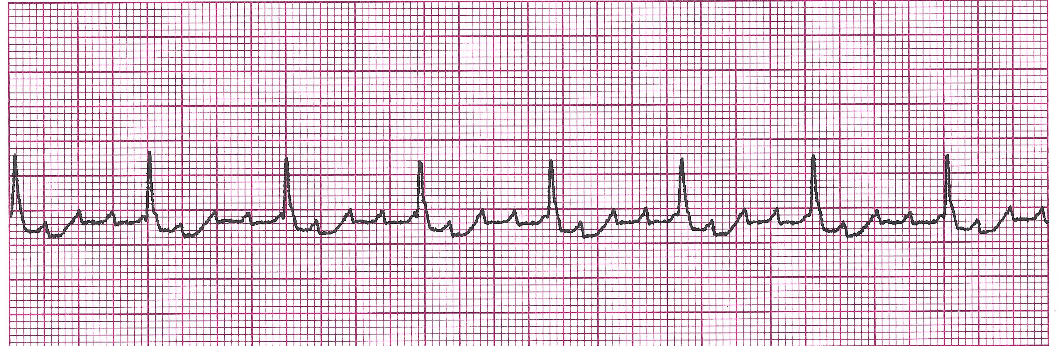
1) Failure to Capture- pacer firing, creating pacer spikes, but heart does not respond (no P wave or QRS after)

2) Failure to Pace- Pacer fails to deliver correct number of stimuli or sometimes any stimulus at all.

3) Competing with patient’s own intrinsic rhythm- can be for two reason and either of these can be bad if the depolarized beat falls near the T wave of the previous beat

pacer competition
Read More
tn_9VentricularRhythms

Ventricular Rhythms

Ventricular Rhythms

What are they?

There are ectopies and escape rhythms. Ectopies arise from an irritable focus in the ventricles. They can be single beats, groups or rums of beats, or entire rhythms. If the ectopic rhythm is faster than the current pacemaker, it will take over the pacemaking of the heart. Ventricular escape rhythms occur when all the pacemaking sites above the ventricles fail and the ventricular pacemaker takes over.

Premature Ventricular Contractions (PVC’s)

Example of PVC

sr with PVC

This is not a rhythm. It is an ectopic beat originating from an irritable ventricular focus. PVC’s usually have the T wave deflecting in the opposite direction of the QRS complex (unlike a normal QRS, T wave). They usually follow by a compensatory pause. This is due to the fact that the SA node did not fire when the PVC did so it waits until the next time it is supposed to fire.

· Rules:

Regularity and rate usually depend on underlying rhythm.

NOTE: PVC’s are usually not counted in the rate determination. However, it may depend on who you ask. The rule is actually to only count the PVC in the rate if it is perfusing, meaning you can feel a pulse with it. Therefore, the best bet is to find, if possible, a 6-second strip of the patient's underlying rhythm without any PVC's to determine the actual heart rate.

Not associated with any P wave, therefore also no PRI

QRS is wide and bizarre: >0.12sec. Will be a different configuration from the other normal QRS complexes in the underlying rhythm.

· Causes: Sometimes there is no apparent cause. Sometimes caused by hypoxia, stress, stimulants, acid-base imbalance, electrolyte imbalance, MI, or CHF.

· Clinical Picture: Can be asymptomatic or may c/o palpitations, racing/fluttering heart, or CP.

· Management: O2, monitor. May use Beta blockers or antiarrhythmics like Lidocaine.

· Interpolated beats

Example of Interpolated PVC’s

PVC_interpolated

Not all PVCs are followed by a pause. If a PVC occurs early enough (especially if the heart rate is slow), it may appear sandwiched in between two normal beats. This is called an interpolated PVC.

· Unifocal vs Multifocal

Example of Unifocal PVC’s

sr unifocal PVC's 2

Unifocal: This means the PVC’s are coming from a single irritable focus in the ventricle. They will be similar in appearance.

Example of Multifocal PVC’s

sr multifocal PVC's

Multifocal: This means the PVC’s originate from different irritable foci in the ventricle. The PVC’s will look differently and possibly deflect in different directions. Multifocal is more serious because it indicates a more irritable ventricle.

· R on T Phenomenon

Example of R on T Phenomenon

R on T into Torsades

This occurs when the R wave of a PVC falls on or near the relative refractory period of the cardiac cycle (the end of the T wave). This is concerning because it can cause the heart to go into a life threatening arrhythmia (as shown in picture above).

· Couplets and Runs

Example of PVC couplet (unifocal)

couplet PVC

Examples of PVC couplet (multifocal)

multifocal couplet

Sometimes PVC’s occur in immediate succession without a normal beat in between. Two in a row are called a couplet. Three or more in a row are called runs. Can represent a very irritable heart and can be serious.

Example of run of PVC’s

(12-15) sr run of 4 vtach

· Grouped Beats

This occurs when the PVC’s fall into a pattern with the surrounding normal beats.

Example of bigeminy

sr PVC big

Bigeminy: Every other beat is a PVC. PVC, one normal beat, PVC, one normal beat, etc.

Example of trigeminy

sr PVC trig 2

Trigeminy: Every third beat is a PVC. PVC, two normal beats, PVC, two normal beats, etc.

Example of quadrigeminy

sr PVC quad 2

Quadrigeminy: Every fourth beat is a PVC. PVC, three normal beats, PVC, three normal beats, etc.

Ventricular Tachycardia

Example of V-tach

vtach

This is a rhythm that originates from an irritable ventricle. Because it is a fast rhythm, it takes over as the pacemaker of the heart. This rhythm can be deadly. Patient may start out ok, but will quickly become unstable and possibly lose their pulse.

· Rules:

Regular or slightly irregular

Rate: Ventricular rate is 150-250bpm. If rate <150bpm, it’s called “Slow VTach”. P waves: None of the QRS’s will have preceding P waves PRI: None QRS: Wide and bizarre, >0.12sec

· Causes: CAD, prior MI, cardiomyopathy, trauma, invasive cardiac procedures, acid-base imbalance, electrolyte imbalance (low Mg, low K, high K), cocaine

· Clinical Picture: Patient will begin to lose consciousness as perfusion decreases. Can quickly become pulseless if they have not already. Can also quickly turn to ventricular fibrillation.

Management:

*Pulse and stable: Lidocaine or Amiodarone

*Pulse and unstable: Use the above meds and cardioversion

*No pulse: CODE BLUE. Start CPR, ACLS. Defibrillate ASAP and continue with ACLS meds.

· Torsades de Pointes

Example of Torsades de Pointes

torsades

Means “twisting of the points”. This is a polymorphic form of V-tach.

Cause: Associated w/ prolonged QT intervals, R on T phenomenon, low Mg, low K. Commonly seen in malnourished individuals and chronic alcoholics. Medication effects that can cause prolonged QT interval such as amiodarone, methadone, lithium, chloroquine, erythromycin, phenothiazines, sotalol, procainamide, quinidine, amitriptyline, fluoxetine, haloperidol, levofloxacin, and sertraline.

Clinical Picture: Patient will have symptoms of low cardiac output. It is hemodynamically unstable and causes a sudden drop in BP, leading to dizziness and syncope. Depending on their cause, many patients revert to normal sinus rhythm within a few seconds, but the rhythm may also persist and possibly deteriorate into ventricular fibrillation.

Example of Torsades de Pointes brought on by R on T Phenomenon

R on T into Torsades

Management: Treat the cause. O2, monitor, get IV access.

IV Magnesium Sulfate. Can quickly deteriorate to ventricular fibrillation.

Ventricular Fibrillation

Example of Ventricular Fibrillation

v fib 2

This is uncoordinated contraction of the ventricles due to multiple irritable foci, making them quiver rather than contract properly and unable to perfuse blood throughout the body. A patient with this rhythm needs immediate ACLS attention. If continues for more than a few seconds will turn into asystole.

· Rules:

Regularity not able to be determined- baseline chaotic.

Rate: indeterminable

P waves: not discernible

PRI: none

QRS: none discernible

· Causes: Increased sympathetic nervous system activity, electrolyte and/or acid-base abnormalities (may need NaHCO3), medication effects, electrocution, trauma, MI.

· Clinical Picture: Clinically dead. Unresponsive, apneic, and pulseless.

· Management: CODE BLUE!! Start CPR, ACLS, and defibrillate ASAP. Use ACLS meds like Lidocaine, Amiodarone.

Idioventricular (Ventricular Escape)

Example of Idioventricular Rhythm

ivr 4

Ventricular escape can be single beats that kick in when the SA or AV nodes fail to fire when they are supposed to, then the SA or AV node kicks back in after the ventricular beat. If it does not kick back in, the patient will proceed to an idioventricular rhythm.

· Rules:

Usually regular

Usually 20-40bpm, but can drop below 20bpm.

No P waves

No PRI

QRS wide >0.12sec.

· Causes: MI, Digoxin toxicity, or other medication effects

· Clinical Picture: Due to the slow rate, patient is usually hypotensive with symptoms of low cardiac output. Can progress to agonal rhythm, then asystole.

· Management: Oxygen, monitor, IV. Atropine, transcutaneous pacing.

· Idioventricular vs Agonal:

Example of Agonal Rhythm

agonal rhythm

Agonal rhythm usually follows idioventricular and represents a dying heart. Usually slower than 20bpm.

· Accelerated Idioventricular

Example of Accelerated Idioventricular Rhythm

AVIR

Usually considered a benign escape rhythm causing little or no hemodynamic instability. Appears when the SA and AV slow too much and disappears when they speed up. Seen commonly after MI’s and with Digoxin toxicity.

Asystole

Example of Asystole

asystole 1

No electrical activity. Just a straight line. However- always check two leads and your patient before determining asystole is indeed occurring. This is because an isoelectric line due to dislodged electrodes can sometimes mimic the appearance of asystole and vice versa. If it is truly asystole: CODE BLUE! Start ACLS immediately with meds like Atropine, Epinephrine, and may try transcutaneous pacing.

Read More

Ventricular Rhythms

Ventricular Rhythms

a. Premature Ventricular Contractions (PVC’s)

Example of PVC

sr with PVC

This is not a rhythm. It is an ectopic beat originating from an irritable area of the ventricle. PVC’s usually have the T wave deflecting in the opposite direction of the QRS complex (unlike a normal QRS, T wave). They are also usually followed by a compensatory pause. This is due to the fact that the SA node did not fire when the PVC did so the SA node waits until the next time it is supposed to fire.

· Rules

Regularity and rate usually depend on underlying rhythm.

NOTE: PVC’s are usually not counted in the rate determination. However, it may depend on who you ask. The rule is actually to only count the PVC in the rate if it is perfusing, meaning you can feel a pulse with it. As a monitor tech, you will likely not be able to do this. Therefore, the best bet is to find, if possible, a 6-second strip of the patient's underlying rhythm without any PVC's to determine the actual heart rate.

Not associated with any P wave, therefore also no PRI

QRS is wide and bizarre: >0.12sec. Will be a different configuration from the other normal QRS complexes in the underlying rhythm.
Concern: Can be concerning- depends on clinical situation and PVC frequency. But many times patients are asymptomatic.

· Interpolated beats

Example of Interpolated PVC’s

PVC_interpolated

Not all PVCs are followed by a pause. If a PVC occurs early enough (especially if the heart rate is slow), it may appear sandwiched in between two normal beats. This is called an interpolated PVC.

· Unifocal vs Multifocal

Example of Unifocal PVC’s

sr unifocal PVC's 2

Unifocal: This means the PVC’s are coming from a single irritable focus in the ventricle. They will be similar in appearance.

Example of Multifocal PVC’s

sr multifocal PVC's

Multifocal: This means the PVC’s originate from different irritable foci in the ventricle. The PVC’s will look differently and possibly deflect in different directions. Multifocal is more serious because it indicates a more irritable ventricle.

· R on T Phenomenon

Example of R on T Phenomenon

R on T into Torsades

This occurs when the R wave of a PVC falls on or near the relative refractory period of the cardiac cycle (the end of the T wave). This is concerning because it can cause the heart to go into a life threatening arrhythmia (as shown in picture above).

· Couplets and Runs

Example of PVC couplet (unifocal)

couplet PVC

Examples of PVC couplet (multifocal)

multifocal couplet

Sometimes PVC’s occur in immediate succession without a normal beat in between. Two in a row are called a couplet. Three or more in a row are called runs. The more PVC’s in a row, the more concerning as it can represent a very irritable heart and turn into a life threatening arrhythmia.

Example of run of PVC’s

(12-15) sr run of 4 vtach

· Grouped Beats

This occurs when the PVC’s fall into a pattern with the surrounding normal beats.

Bigeminy: Every other beat is a PVC. PVC, one normal beat, PVC, one normal beat, etc.

Example of bigeminy

sr PVC big

Trigeminy: Every third beat is a PVC. PVC, two normal beats, PVC, two normal beats, etc.

Example of trigeminy

sr PVC trig 2

Quadrigeminy: Every fourth beat is a PVC. PVC, three normal beats, PVC, three normal beats, etc.

Example of quadrigeminy

sr PVC quad 2

c. Ventricular Tachycardia

Example of V-tach

vtach

This is a rhythm that originates from an irritable ventricle. This rhythm can be deadly. The patient may start out ok, but will quickly become unstable and possibly lose their pulse.

· Rules:

Regular or slightly irregular

Rate: Ventricular rate is 150-250bpm. If rate <150bpm, it’s called “Slow VTach”. P waves: None of the QRS’s will have preceding P waves PRI: None QRS: Wide and bizarre, >0.12sec

Concern: EMERGENCY SITUATION- CAN TURN INTO A CODE BLUE!!

· Torsades de Pointes

Example of Torsades de Pointes

torsades

Means “twisting of the points”. This is a polymorphic form of V-tach.

Cause: Associated w/ prolonged QT intervals, R on T phenomenon.

Example of Torsades de Pointes brought on by R on T Phenomenon

R on T into Torsades

Concern: EMERGENCY SITUATION- CAN TURN INTO A CODE BLUE!!

d. Ventricular Fibrillation

Example of Ventricular Fibrillation

v fib 2

This represents very irritable ventricles, making them quiver rather than contract properly and unable to perfuse blood throughout the body. A patient with this rhythm needs immediate ACLS attention. If continues for more than a few seconds will turn into asystole.

Rules:

Regularity not able to be determined- baseline chaotic.

Rate: indeterminable

P waves: not discernible

PRI: none

QRS: none discernible

Concern: EMERGENCY SITUATION! CODE BLUE!

e. Idioventricular (Ventricular Escape)

ivr 4

Example of Idioventricular Rhythm

Ventricular escape can be single beats that kick in when the SA or AV nodes fail to fire when they are supposed to, then the SA or AV node kicks back in after the ventricular beat. If it does not kick back in, the patient will proceed to an idioventricular rhythm.

· Rules:

Usually regular

Usually 20-40bpm, but can drop below 20bpm.

No P waves

No PRI

QRS wide >0.12sec.

Concern: Very concerning- needs immediate attention

· Idioventricular vs Agonal:

Example of Agonal Rhythm

agonal rhythm

Agonal rhythm usually follows idioventricular and represents a dying heart. Usually slower than 20bpm.

· Accelerated Idioventricular

Example of Accelerated Idioventricular Rhythm

AVIR

Usually considered a benign escape rhythm causing little or no hemodynamic instability. Appears when the SA and AV slow too much and disappears when they speed up.

Concern: Can be. Depends on clinical situation.

f. Asystole

Example of Asystole

asystole 1

No electrical activity. Just a straight line. However- always check two leads and your patient before determining asystole is indeed occurring. This is because an isoelectric line due to dislodged electrodes can sometimes mimic the appearance of asystole and vice versa. If it is truly asystole: CODE BLUE!

STOP! TIME FOR PRACTICE STRIPS ON VENTRICULAR RHYTHMS!

Read More
tn_8AtrioventricularBlocks

Atrioventricular Blocks

1. Atrioventricular Blocks

a. What are they?

Cardiac conduction system

These conditions do not come from the AV node. They are a result of an impulse from somewhere above the AV node having trouble getting through the AV junction or the top of the Bundle of His. They are categorized according to the severity of obstruction in the AV junction.

b. First Degree AV block

Example of 1st degree AV block

(12-2) 1st HB 1

Not a true block because each impulse is conducted. It just takes longer than usual.The block is only partial. It is least serious of all the blocks.

REMEMBER: THIS IS NOT A RHYTHM BY ITSELF. IT IS A CONDITION SUPERIMPOSED ON AN EXITING RHYTHM.

· Rules:

Regularity and rate depend on the underlying rhythm

P waves are upright, uniform, and before every QRS

PRI: >0.20sec and constant

QRS: <0.12sec

· Causes: myocarditis, MI, hypothyroid, cardiomyopathies, Digoxin toxicity, anoxia, increased vagal tone, electrolyte imbalances, medication effects like Ca channel blockers, Beta-blockers, cardiac glycosides

· Clinical Picture: Usually asymptomatic, but can lead to a more serious AV block.

· Management: Treat the cause. O2, monitor. Can lead to a more serious AV block.

c. Second Degree AV block MobitzType I (Wenckebach)

Example of 2nd degree AV block Type I (Wenckebach)

(12-32)2nd HB Wenckebach 2

This block is more serious that first degree because not all impulses get conducted through the AV node. The only differences between Second Degree Type I and Type II are the PRI and the number of impulses allowed through the AV node. You will usually see more impulses being conducted with Type I.

· Rules:

Regularly Irregular (Pattern)

Ventricular rate will be slightly slower than usual due to the dropped beat.

P waves upright, uniform, but not always followed by a QRS

PRI gets progressively longer until one P wave is not followed by a QRS (a blocked beat). After this “blocked beat”, the cycle starts again.

QRS: <0.12

· Causes: Medication effects like Digitalis, Beta-blockers, Ca channel blockers, increased parasympathetic tone, myocardial ischemia at or near the AV node, inferior MI

· Clinical Picture: Usually asymptomatic

· Management: Treat the cause. O2. Monitor. Can lead to a more serious AV block. Can also use Atropine, Epinephrine, or transcutaneous pacing if patient becomes unstable.

d. Second Degree AV block Mobitz Type II (Classic)

Example of 2nd degree AV block Type II (Classic)

(12-17) 2nd HB Classic 5

· Rules: Regularity may vary depending on how many and which impulses are allowed through.If the conduction ratio is consistent (i.e- every other P wave is not followed by a QRS), then the R-R will be constant and regular. If the conduction ratio varies (i.e. two P waves have QRS then one does not, then one P wave has QRS, etc), the R-R will be irregular.

Ventricular rate will usually be bradycardic due to all the blocked impulses

P waves are upright and uniform. Not all are followed by QRS.

PRI: Constant throughout conducted beats. However, it can be >0.20.

QRS <0.12, however can also be wider depending on the physical location of the block. The lower the block below the AV node, the more likely the QRS will be wider.

· Causes: myocardial ischemia or infarct, myocarditis, Ca channel blockers, Amiodarone, Digoxin

· Clinical Picture: Can by asymptomatic but , if the rate is slow, patient may show signs of decreased cardiac output, SOB, CHF.

· Management: Treat the cause, give O2, establish IV site if patient does not have one already, and MONITOR as this rhythm commonly transitions to complete heart block. Can use Atropine, Epinephrine, and transcutaneous pacing if patient becomes unstable.

e. Third Degree AV block (Complete)

Example of 3rd degree AV block

Third-degree-heart-block

Third degree heart block occurs when there is a complete block at the AV junction. No impulses conducted from the SA, atria, and sometimes the AV junction can get through to the ventricles. For this reason a lower escape pacemaker (either the lower AV junction or ventricles) kicks in and conducts impulses for the ventricles while the atrial pacemaker (SA, AV node, or atria) conducts impulses for the atria. Because they have different pacemaker sites, the atria and ventricles are beating independently of each other and there is no relationship between the two. Therefore, the contraction of each chamber is not sequenced correctly and the heart cannot fill with blood adequately. Moreover, because the ventricular pacemaker only beats at 20-40bpm, the patient may be very bradycardic and hemodynamically unstable.

· Rules:

P-P intervals and R-R intervals are regular and constant.

Ventricular rate depends on the controlling focus. If junctional: 40-60bpm, if ventricular: 20-40bpm.

P waves will be upright and uniform. Always more P waves than QRS’s and no consistent relationship between P waves and QRS.

PRI: Does not exist since there is no conduction to the ventricles. No relationship exists.

QRS: Depends on the controlling focus. If junctional, QRS will be <0.12, if ventricular, may measure 0.12 or greater.

· Causes: Commonly associated with MI. Other causes include ischemic heart disease, excessive vagal tone, Digoxin toxicity, Ca channel blockers, Beta blockers, electrolyte imbalance, myocarditis.

· Clinical Picture: Usually shows signs and symptoms of low cardiac output. May become unstable.

· Management: Oxygen, Monitor, establish IV site, if patient does not have one. Transcutaneous pacing. Treat the cause, if possible.

Read More

Atrioventricular Blocks

. Atrioventricular Blocks

a. What are they?

Cardiac conduction system

These conditions do not come from the AV node. They are a result of an impulse from somewhere above the AV node having trouble getting through the AV junction or the top of the Bundle of His. They are categorized according to the severity of obstruction in the AV junction.

b. First Degree AV block

Example of 1st degree AV block

(12-2) 1st HB 1

Not a true block because each impulse is conducted. It just takes longer than usual. The block is only partial. It is least serious of all the blocks. REMEMBER: THIS IS NOT A RHYTHM BY ITSELF. IT IS A CONDITION SUPERIMPOSED ON AN EXITING RHYTHM.

· Rules:

Regularity and rate depend on the underlying rhythm

P waves are upright, uniform, and before every QRS

PRI: >0.20sec and constant

QRS: <0.12sec

· Causes: myocarditis, MI, hypothyroid, cardiomyopathies, Digoxin toxicity, anoxia, increased vagal tone, electrolyte imbalances, medication effects like Ca channel blockers, Beta-blockers, cardiac glycosides

· Clinical Picture: Usually asymptomatic, but can lead to a more serious AV block.

· Management: Treat the cause. O2, monitor. Can lead to a more serious AV block.

c. Second Degree AV block MobitzType I (Wenckebach)

Example of 2nd degree AV block Type I (Wenckebach)

(12-32)2nd HB Wenckebach 2

This block is more serious that first degree because not all impulses get conducted through the AV node. The only differences between Second Degree Type I and Type II are the PRI and the number of impulses allowed through the AV node. You will usually see more impulses being conducted with Type I.

· Rules:

Regularly Irregular (Pattern)

Ventricular rate will be slightly slower than usual due to the dropped beat.

P waves upright, uniform, but not always followed by a QRS

PRI gets progressively longer until one P wave is not followed by a QRS (a blocked beat). After this “blocked beat”, the cycle starts again.

QRS: <0.12

· Causes: Medication effects like Digitalis, Beta-blockers, Ca channel blockers, increased parasympathetic tone, myocardial ischemia at or near the AV node, inferior MI

· Clinical Picture: Usually asymptomatic

· Management: Treat the cause. O2. Monitor. Can lead to a more serious AV block. Can also use Atropine, Epinephrine, or transcutaneous pacing if patient becomes unstable.

d. Second Degree AV block Mobitz Type II (Classic)

Example of 2nd degree AV block Type II (Classic)

(12-17) 2nd HB Classic 5

· Rules:

Regularity may vary depending on how many and which impulses are allowed through. If the conduction ratio is consistent (i.e- every other P wave is not followed by a QRS), then the R-R will be constant and regular. If the conduction ratio varies (i.e. two P waves have QRS then one does not, then one P wave has QRS, etc), the R-R will be irregular.

Ventricular rate will usually be bradycardic due to all the blocked impulses

P waves are upright and uniform. Not all are followed by QRS.

PRI: Constant throughout conducted beats. However, it can be >0.20.

QRS <0.12, however can also be wider depending on the physical location of the block. The lower the block below the AV node, the more likely the QRS will be wider.

· Causes: myocardial ischemia or infarct, myocarditis, Ca channel blockers, Amiodarone, Digoxin

· Clinical Picture: Can by asymptomatic but , if the rate is slow, patient may show signs of decreased cardiac output, SOB, CHF.

· Management: Treat the cause, give O2, establish IV site if patient does not have one already, and MONITOR as this rhythm commonly transitions to complete heart block. Can use Atropine, Epinephrine, and transcutaneous pacing if patient becomes unstable.

e. Third Degree AV block (Complete)

Example of 3rd degree AV block

Third-degree-heart-block

Third degree heart block occurs when there is a complete block at the AV junction. No impulses conducted from the SA, atria, and sometimes the AV junction can get through to the ventricles. For this reason a lower escape pacemaker (either the lower AV junction or ventricles) kicks in and conducts impulses for the ventricles while the atrial pacemaker (SA, AV node, or atria) conducts impulses for the atria. Because they have different pacemaker sites, the atria and ventricles are beating independently of each other and there is no relationship between the two. Therefore, the contraction of each chamber is not sequenced correctly and the heart cannot fill with blood adequately. Moreover, because the ventricular pacemaker only beats at 20-40bpm, the patient may be very bradycardic and hemodynamically unstable.

· Rules:

P-P intervals and R-R intervals are regular and constant.

Ventricular rate depends on the controlling focus. If junctional: 40-60bpm, if ventricular: 20-40bpm.

P waves will be upright and uniform. Always more P waves than QRS’s and no consistent relationship between P waves and QRS.

PRI: Does not exist since there is no conduction to the ventricles. No relationship exists.

QRS: Depends on the controlling focus. If junctional, QRS will be <0.12, if ventricular, may measure 0.12 or greater.

· Causes: Commonly associated with MI. Other causes include ischemic heart disease, excessive vagal tone, Digoxin toxicity, Ca channel blockers, Beta blockers, electrolyte imbalance, myocarditis.

· Clinical Picture: Usually shows signs and symptoms of low cardiac output. May become unstable.

· Management: Oxygen, Monitor, establish IV site, if patient does not have one. Transcutaneous pacing. Treat the cause, if possible.

STOP! TIME FOR PRACTICE STRIPS ON AV BLOCKS!

Read More
tn_7JunctionalRhythms

Junctional Rhythms and SVT

1. Junctional Rhythms

Cardiac conduction system

Cardiac conduction system

Conduction works differently with junctional rhythms. The impulse comes from the AV node which is anatomically in the middle of the heart. In order for the atria and ventricles to get depolarized, the impulse needs to travel in both directions meaning the atria and ventricles usually get

depolarized at about the same time. When the impulse travels backward todepolarize the atria, it is called “retrograde” conduction. This backwards impulse causes the P wave to look different, if it can be seen at all, on the EKG.

a. P waves in Junctional Rhythms

Inverted P waves:

Inverted P wave example

In Lead II, remember, the “camera” is at the apex of the heart. If the impulse starts at the AV junction and has to travel in both directions, the P wave will be a negative deflection because the current is flowing away from the “camera”. The QRS will be positive as usual.

. Hidden P waves:

Junctional

Because the depolarization of the atria occurs at the same time as the ventricles, the P wave may not be seen at all because it is hidden by the QRS.

· Late P waves:

P wave after QRS (bem.fi.com)

The impulse may also reach and depolarize the ventricles before the atria. In this instance, the P wave comes after the QRS.

b. Premature Junctional Contractions (PJC’s)

Example of PJC

SR with PJC (6.10) circled

These are single ectopic beats in a rhythm. Occurs when an irritable focus in the junction stimulates a depolarization and interrupts the underlying rhythm for a single beat. Appears similar to PAC except the P wave will look different (see P waves section above).

· Rules:

Basically irregular due to interruption

Rate will depend upon underlying rhythm

P wave: will appear inverted, and come before, during, or after QRS

PRI: If P wave precedes QRS, will be usually less than 0.12sec

QRS: <0.12sec

· Causes: Nicotine, hypoxia, dig toxicity, caffeine, MI, stress, alcohol, infections, heart failure, mental or physical fatigue, hypomagnesemia, hypokalemia, or can occur in healthy hearts

· Clinical Picture: May feel palpitations or skipped beats

· Management: Usually none needed

c. Junctional Escape

Example of Junctional Escape Rhythm

Junctional

When higher pacemaker sites fail, the AV junction is left with pacemaking responsibility.

· Rules:

Regular

Rate 40-60bpm

P wave: Inverted and can come before, during, or after QRS

PRI: If P wave precedes QRS, will be

QRS: <0.12sec

· Cause: Severe sinus bradycardia, vasovagal stimulation, hyperkalemia, drug effects like Beta-blockers, Ca channel blockers, Dig toxicity

· Clinical Picture: Slow pulse, may have symptoms of decreased cardiac output like, dizziness, fatigue, short of breath, syncope, but may be asymptomatic.

· Management: O2, monitor IV. Treat the cause. If symptomatic, drug therapy like atropine or pacemaker may be needed.

d. Accelerated Junctional Rhythm

Example of Accelerated Junctional Rhythm

accelerated junctional 1 (6.8)

Occurs when an irritable site at the AV junction sends impulses at a faster rate than the SA node or other atrial pacemaker sites and takes over pacemaker responsibility of the heart.

· Rules:

Regular

Rate 60-100bpm

P waves are inverted and can come before, during, or after the QRS

PRI: If P wave precedes the QRS, it will be

QRS: <0.12sec

· Causes: MI, open heart surgery, myocarditis, Digoxin toxicity

· Clinical Picture: Usually asymptomatic

· Management: O2, monitor, IV. Treat cause.

e. Junctional Tachyicardia

Example of Junctional Tachycardia

junctional tachycardia (6.17)

· Rules: Regular

Rate 100-180bpm (NOTE: most of the time,

when >150, it may be hard to tell if rhythm is sinus, atrial, or junctional in origin, so new call it SVT).

P wave: Inverted and can come before, during, or after QRS

PRI: If P wave precedes QRS will be

QRS: <0.12sec

· Causes: MI, open heart surgery, myocarditis, Digoxin toxicity

· Clinical Picture: Fast pulse, may shows signs of decreased cardiac output, palpitations.

· Management: O2, monitor, IV. Vasovagal maneuvers, carotid massage, drug therapy such as Adenosine, Ca channel blockers, Beta-blockers, antiarrythmics, or cardioversion

2. Supraventricular Tachycardias (SVT)

a. What are they?

Example of SVT

svt

A category of several regular tachyarrhythmias that can’t be identified further due to indistinguishable P waves, rate 150-250bpm. Is usually one of four rhythms:sinus tach, atrial tach, atrial flutter, or junctional tach.

b. P waves in SVT

Not discernible due to the fast rate of the rhythm.

STOP! TIME FOR PRACTICE STRIPS ON JUNCTIONAL RHYTHMS / SVT

Read More

Junctional Rhythms and SVT

1. Junctional Rhythms

Cardiac conduction system

Cardiac conduction system

Conduction works differently with junctional rhythms. The impulse comes from the AV node which is anatomically in the middle of the heart. In order for the atria and ventricles to get depolarized, the impulse needs to travel in both directions meaning the atria and ventricles usually get depolarized at about the same time. When the impulse travels backward to depolarize the atria, it is called “retrograde” conduction. This backwards impulse causes the P wave to look different, if it can be seen at all, on the EKG.

a. P waves in Junctional Rhythms

· Inverted P waves:

Inverted P wave example

In Lead II, remember, the “camera” is at the apex (bottom) of the heart. If the impulse starts at the AV junction and has to travel in both directions, the P wave will be a negative deflection because the current is flowing away from the “camera”. The QRS will be positive as usual.

· Hidden P waves:

Junctional

Because the depolarization of the atria occurs at the same time as the ventricles, the P wave may not be seen at all because it is hidden by the QRS.

· Late P waves:

P wave after QRS (bem.fi.com)

The impulse may also reach and depolarize the ventricles before the atria. In this instance, the P wave comes after the QRS.

b. Premature Junctional Contractions (PJC’s)

Example of PJC

SR with PJC (6.10) circled

These are single ectopic beats in a rhythm. Occurs when an irritable focus in the junction stimulates a depolarization and interrupts the underlying rhythm for a single beat. Appears similar to PAC except the P wave will look different (see P waves section above).

· Rules:

Basically irregular due to interruption

Rate will depend upon underlying rhythm

P wave: will appear inverted, and come before, during, or after QRS

PRI: If P wave precedes QRS, will be usually less than 0.12sec

QRS: <0.12sec

· Concern: Can be concerning. Depends on clinical situation and frequency.

c. Junctional Escape

Example of Junctional Escape Rhythm

Junctional

When higher pacemaker sites fail, the AV junction is left with pacemaking responsibility.

· Rules:

Regular

Rate 40-60bpm

P wave: Inverted and can come before, during, or after QRS

PRI: If P wave precedes QRS, will be

QRS: <0.12sec

Concern: Can be concerning. Depends on clinical situation. The slower the rate, the more concern.

d. Accelerated Junctional Rhythm

Example of Accelerated Junctional Rhythm

accelerated junctional 1 (6.8)

Occurs when an irritable site at the AV junction sends impulses at a faster rate than the SA node or other atrial pacemaker sites and takes over pacemaker responsibility of the heart.

· Rules:

Regular

Rate 60-100bpm

P waves are inverted and can come before, during, or after the QRS

PRI: If P wave precedes the QRS, it will be

QRS: <0.12sec

Concern: Can be concerning. Depends on clinical situation

e. Junctional Tachyicardia

Example of Junctional Tachycardia

junctional tachycardia (6.17)

· Rules:

Regular

Rate 100-180bpm (NOTE: most of the time, when >150, it may be hard to tell if rhythm is sinus, atrial, or junctional in origin, so new call it SVT).

P wave: Inverted and can come before, during, or after QRS

PRI: If P wave precedes QRS will be

QRS: <0.12sec

Concern: Concerning- needs attention

2. Supraventricular Tachycardias (SVT)

a. What are they?

A category of several regular tachyarrhythmias that can’t be identified further due to indistinguishable P waves, rate 150-250bpm. Is usually one of four rhythms:sinus tach, atrial tach, atrial flutter, or junctional tach.

Example of SVT

svt

b. P waves in SVT

Not discernible due to the fast rate of the rhythm.

STOP! TIME FOR PRACTICE STRIPS ON JUNCTIONAL RHYTHMS / SVT

Read More
tn_6AtrialRhythms

Atrial Rhythms

Atrial Rhythms

Cardiac conduction system

Cardiac conduction system

These are rhythms that originate somewhere in the atria other than the SA node. Causes include loss of SA node capability and atrial irritability. P waves in this category will have a different appearance than those beats generated from the SA node (can be notched, flattened, peaked, or sawtooth).

Wandering atrial pacemaker

Example Wandering atrial pacemaker

WAP (9.128)

· Rules: R-R slightly irregular ,

Rate 60-100bpm ,

P wave appearance changes with each beat ,

PRI varies but is usually

· Causes: Can be normal- seen in very young, very old, or athletic patients

· Clinical Picture: Usually asymptomatic

· Management: None needed

Premature Atrial Contractions (PAC’s)

It is a type of atrial ectopy caused by irritability. It is a single beat. It is important that you always identify the underlying rhythm and locate the ectopies throughout that rhythm.

Example of PAC

SR with PAC (5.3) (circled)

· Rules: Rhythm will appear irregular due to the ectopic beats interrupting.

Rate depends on underlying rhythm

P wave of the PAC will look different from the rest of the strip. It usually has a QRS following but can be blocked.

PRI 0.12-0.20sec (PAC’s PRI may be longer or different from the others)

QRS <0.12sec

· Causes: fatigue, hypoxia, Digoxin toxicity, caffeine, ischemia, CHF, or ETOH

· Clinical Picture: irregular pulse, but usually asymptomatic

· Management: Treat the cause. Monitor as it can lead to atrial tachyarrythmias. Oxygen, monitor, IV.

Atrial Tachycardia (AT)

Because of its fast rate and supraventricular origin, atrial tachycardia usually gets interpreted as SVT (See SVT in next section)

· Rules:

Regular

150-250bpm

P wave is atrial in origin (not sinus) and may be lost or hidden in T wave due to the fast rate

PRI 0.12-0.20sec

QRS <0.12sec

· Causes: Excessive amount of catecholamines like epinephrine, norepinephrine, and dopamine, or Digoxin toxicity

· Clinical Picture: Rapid, regular pulse. May show symptoms of decreased cardiac output like, dizziness, fatigue, short of breath, syncope. May have chest pain or palpitations

· Management: O2, monitor, IV. Vasovagal maneuvers, carotid massage, drug therapy like Ca channel blockers, Beta-blockers, antiarrhythmics, or cardioversion.

Atrial Flutter (A-Flutter)

Example of Atrial flutter

(12-16) aflutter (5.41)

· Rules: Usually regular but can be irregular

Atrial rate is usually 250-350bpm, ventricular rate can vary

P waves are upright and sawtooth

PRI –not applicable

QRS <0.12sec

· Causes: CAD,Rheumatic heart disease, hyperthyroid, MI

· Clinical Picture: If there is a rapid ventricular response- patient may have symptoms of decreased cardiac output like, dizziness, fatigue, short of breath, syncope and palpitations. Can lead to CHF.

· Management: Oxygen, monitor, IV. Treat the cause. Anticoagulant therapy is initiated due to increased risk of clots (the rapid atrial contraction causes turbulence in the blood which the body thinks is an injury- this causes platelets to collect at the site of the turbulence causing clots). If there is RVR, drug therapy such as Ca channel blockers, beta-blockers, and antiarrhythmics like Digoxin, Corvert, or Amiodarone can be used. Atrial pacing and cardioversion are options if patient becomes unstable.

Atrial Fibrillation (A-Fib)

Example of A-Fib

afib (5.29)

· Rules: Totally irregular.

If ventricular rate 100 “Rapid ventricular response”

No true P waves are produced in this rhythm, just fibrillatory waves

PRI unmeasurable

QRS <0.12sec

· Causes: MI, Digoxin toxicity

· Clinical Picture: Irregular pulse, may have symptoms of decreased cardiac output like, dizziness, fatigue, short of breath, syncope and palpitations. Can lead to CHF.

· Management: Oxygen, monitor, IV. Treat the cause. Anticoagulant therapy is initiated due to increased risk of clots (the rapid atrial contraction causes turbulence in the blood which the body thinks is an injury- this causes platelets to collect at the site of the turbulence causing clots). If there is RVR, drug therapy such as Ca channel blockers, beta-blockers, and antiarrhythmics like Digoxin, Corvert, or Amiodarone can be used. Atrial pacing and cardioversion are options if patient becomes unstable.

STOP! TIME FOR PRACTICE STRIPS ON VARIOUS ATRIAL RHYTHMS

Read More

Atrial Rhythms

Atrial Rhythms

Cardiac conduction system

Cardiac conduction system

These are rhythms that originate somewhere in the atria other than the SA node. Causes include loss of SA node capability and atrial irritability. P waves in this category will have a different appearance than those beats generated from the SA node (can be notched, flattened, peaked, or sawtooth).

a. Wandering atrial pacemaker

Wandering atrial pacemaker

WAP (9.128)

· Rules:

R-R slightly irregular

Rate 60-100bpm

P wave appearance changes with each beat

PRI varies but is usually <0.20sec

QRS <0.12sec

Concern: Usually none

· b. Premature Atrial Contractions (PAC’s)

It is a type of atrial ectopy caused by irritability. It is a single beat. It is important that you always identify the underlying rhythm and locate the ectopies throughout that rhythm.

Example of PAC

SR with PAC (5.3) (circled)

· Rules:

Rhythm will appear irregular due to the ectopic beats interrupting.

Rate depends on underlying rhythm

P wave of the PAC will look different from the rest of the strip. It usually has a QRS following but can be blocked.

PRI 0.12-0.20sec (PAC’s PRI may be longer or different from the others)

QRS <0.12sec

Concern: Can be concerning. Depends on clinical situation. Depends on frequency.

c. Atrial Tachycardia (AT)

Because of its fast rate and supraventricular origin, atrial tachycardia usually gets interpreted as SVT (See SVT in next section)

Rules:

Regular

150-250bpm

P wave is atrial in origin (not sinus) and may be lost or hidden in T wave due to the fast rate

PRI 0.12-0.20sec

QRS <0.12sec

Concern: Yes- needs attention.

d. Atrial Flutter (A-Flutter)

Example of Atrial flutter

(12-16) aflutter (5.41)

· Rules:

Usually regular but can be irregular

Atrial rate is usually 250-350bpm, ventricular rate can vary

P waves are upright and sawtooth

PRI –not applicable

QRS <0.12sec

Concern: Can be concerning. Depends on clinical situation

e. Atrial Fibrillation (A-Fib)

Example of A-Fib

afib (5.29)

· Rules:

Totally irregular.

If ventricular rate 100 “Rapid ventricular response”

No true P waves are produced in this rhythm, just fibrillatory waves

PRI unmeasurable

QRS <0.12sec

Concern: Can be concerning. Depends on clinical situation

STOP! TIME FOR PRACTICE STRIPS ON VARIOUS ATRIAL RHYTHMS

Read More
tn_5SinusRhythms

Sinus Rhythms

Sinus Rhythms

Normal Sinus Rhythm

Examples of NSR

Click to enlarge

· Rules: 

Regular

60-100bpm

P wave is uniform, upright ,and there is one in front of every QRS

PRI is 0.12-0.20 and constant

QRS <0.12

· Causes: Normal

· Clinical Picture: Asymptomatic

· Management: None needed

Sinus Bradycardia
Example of Sinus bradycardia

Click to enlarge

· Rules: Regular

<60bpm

P wave uniform, upright, and one before every QRS

PRI 0.12-0.20sec and constant

QRS <0.12sec

· Causes: Sleep, hypothyroid, drug effects like beta-blockers, Ca+ channel blockers, and antiarrythmics, MI, increased ICP, increased vagal tone (i.e.athletes).

· Clinical Picture: Often asymptomatic, but may show symptoms of decreased cardiac output like, dizziness, fatigue, short of breath, syncope

· Management: If symptomatic, may give drugs to increase HR, apply pacemaker, etc. Also use O2, monitor, IV.

Sinus Tachycardia

Example of Sinus tachycardia

(12-41)ST (4.23)

· Rules: Regular ,

<100bpm ,

P wave same as above ,

PRI 0.12-0.20sec, constant ,

QRS <0.12sec

· Causes: Stress, anxiety, dehydration, pulmonary embolus, fever, exercise, anemia, drug effects (caffeine, cocaine, nicotine), shock, MI, hyperthyroid

· Clinical Picture: Often asymptomatic, but depends on cause. May have palpitations, SOB, chest pain

· Management: Usually should try to treat the cause. Also Use O2, monitor, IV.

Sinus Arrythmia

Example of Sinus Arrhythmia

sinus arrythmia

Also called “Respiratory Sinus Arrhythmia”. Occurs when your heart rate cycles with your breathing. When you breathe in, your heart rate speeds up slightly. When you breathe out, your heart rate slows back down.

· Rules: R-R intervals vary; change with patient’s respirations ,

Rate usually 60-100bpm, but can be slower ,

P wave same as above , PRI 0.12-0.20sec, constant ,

QRS <0.12sec

· Causes: Usually a normal phenomenon. Common in children, young adults, and athletes.

· Clinical Picture: Irregular pulse but usually asymptomatic

· Management: Usually none needed

Sinus pauses and sinus arrest

Caused when the sinus node fails to generate an electrical impulse to stimulate the heart to beat. Many times both of these categories are lumped into one called “Pauses” when interpreting rhythms.

1) Sinus Pause/Sinus block

A sinus pause is followed by a delayed, next sinus beat only.

Example of Sinus pause/Sinus block

sinoatrial block

· Rules: Irregular due to pause. Pause must be followed by a sinus beat.

Length of the pause is usually the same as or a multiple of the distance between two other P-P intervals.

Rate is usually normal but depends on the underlying rhythm.

P waves are uniform, upright, and before every QRS.

PRI 0.12-0.20sec, constant

QRS <0.12sec

· Causes: acute MI, medication effects like Digitalis, and other antiarrhythmics, CAD, myocarditis, CHF, increased vagal tone

· Clinical Picture: May show symptoms of decreased cardiac output like, dizziness, fatigue, short of breath, syncope

· Management: Usually treat the cause. Use O2, monitor, IV.

2) Sinus Arrest

A pause, however, many times is followed by an escape beat. This is called sinus arrest as the sinus node is taking too long to beat again needs help from the AV node.

NOTE: Patients with sinus arrest may have a history of syncope, near syncope, and/or falls.

sinus arrest

· Rules: Irregular due to pause.

Pauses will vary in length and are usually more than one PQRST complex long.

Rate varies depending on underlying rhythm.

P waves uniform, upright, and one before every QRS

PRI 0.12-0.20sec, constant

QRS <0.12sec

· Causes: hypoxia, MI, high potassium, Digoxin toxicity, medication effects like Beta-blockers and Ca+ channel blockers

· Clinical Picture: May show symptoms of decreased cardiac output like, dizziness, fatigue, short of breath, syncope

· Management: Usually treat the cause. Use O2, monitor, IV.

Facts

JUST WHAT IS "SICK SINUS SYNDROME"??

It occurs as a bradycardia with episodes of sinus arrest, sinoatrial block, and atrial fibrillation. Caused by partial destruction of the SA node from things like an MI. Can cause prolonged pauses in heart rate and rhythm and syncope. Many patients with sick sinus syndrome end up receiving a pacemaker.

STOP! TIME FOR PRACTICE STRIPS ON VARIOUS SINUS RHYTHMS

Read More